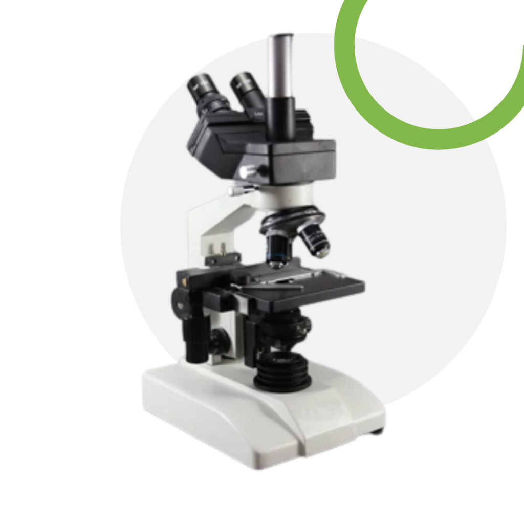
Trinocular Microscope
WLS-TCM
A trinocular microscope has two eyepieces like a binocular microscope and an additional third eye tube for connecting a microscope camera. They are therefore a binocular with a moving prism assembly in which light is either directed to the binocular assembly of the microscope or to the camera.

About the product
A trinocular microscope has two eyepieces like a binocular microscope and an additional third eye tube for connecting a microscope camera. They are therefore a binocular with a moving prism assembly in which light is either directed to the binocular assembly of the microscope or to the camera.
The best models of this microscope will have at least three positions, allowing 100 percent of light to the binocular, 80 percent to camera and 20 percent to the binocular or simply a 100 percent to the camera.
One of the biggest advantages of this microscope (three position trinocular) is its versatility. For instance, for brightfield photographic purposes, the 20% visual- 80% photo system would be the ideal choice.
The amount of light provided is ideal allowing the camera a very short exposure time, which in turn ensures that the specimen is seen in real time. This makes photography a lot easier. On the other hand, fluorescence photography requires that the trinocular be set at 100% to the binocular given that this type of photographing requires a lot of light.
Features & Benefits
- Ones prepared for observation; most specimens will start undergoing changes over a period of time. For the most part, they cannot be preserved and viewed long term.
- A trinocular microscope allows for the user to not only take pictures, but also record videos, which can be saved for future references. This is also a big advantage given that such still images and videos can give more details after repeated observations. In a clinical setting, this is particularly beneficial given that health care professionals can digitally share the images/videos with other professionals for consultation and more analysis.
- For teaching purposes (e.g., microscopy), a trinocular microscope also presents an advantage given that an instructor can show the students what he/she is looking at, or observe how the student is using the microscope. On the other hand, it can also be used for presentation purposes either in academic of professional settings, allowing others to participate in the viewing of the specimen.
- For younger students, it provides an opportunity to record videos and take photos, which they can then compare with other images in books, and learn more about the different parts of cells or other organisms they observed.
Product Specifications
| OPTICAL SYSTEM | Infinity corrected optical system |
| STAND | Stable & sturdy shaped stand welk-contoured modular base corrosion resistant paint and heat resistant pads. |
| VIEWING HEAD | Siedentopf binocular/Trinocular tube. Inclined at 30o rotatable through 360o with IPD 55-75 mm. Anti fungus treated. Diopter adjustment on one tube. |
| EYEPIECES | Eyepiece WF 10x FN 20 paired with eye guards, anti-fungus treated. |
| CAMERA | · Image Sensor: ½ 5" CMOS 3.0MP. |
| · Resolution: 2048 X 1536 (3.0M Pixels) | |
| · Frame Rate: 3fps@2048x1536; 30 fps@640 x480. | |
| · Output Interface: USB-2.0/3.0, | |
| · Software: Preview. static capture, video capture. | |
| · System Required: Microsoft windows 2000, XP SP2JSP3, VistaUSB2.0 Port. | |
| · Application: High resolution microscope imaging, | |
| · Mount: C/CS mount | |
| · Mounting Tube: 23mm. | |
| · Camera size: 60mm diameter | |
| · Image File Type: JPEG. PNG, 13MP, TIF. | |
| NOSEPIECE | Low friction & fully parfocal Reverse angle quadruple Revolving nosepiece (Ball bearing type) with click stop & rubber grip. |
| OBJECTIVES | Eco Infinity Plan Achromatic, Anti fungus treated objectives: 4x0.10, 10x0.25, 40x (SL)/0.65, 100x (SL, OIL)/1.25. |
| FOCUSING | Ergonomic low position co-axial coarse and fine focusing system on ball bearing guide ways. |
| STAGE | Double layer graduated mechanical rectangular stage size 140 x 132mm wire cross travel 75 (X) x 50 (Y) mm on ball bearing co-axial contras spring dip specimen holder. |
| CONDENSER | Movable ABBE condenser, with aspheric lens NA 1.25, daylight blue filter & Iris diaphragm. Centrable |
| ILLUMINATION | Super Bright White LED cold Light Optional 6V 20W Halogen Lamp |
| POWER | Universal Power Supply 100v to 240v AC 5060 Hertz. |
| PACKAGING CONTENT | The microscope is packed inside a STYROFOAM box with dust cover |
Book Your Equipment Demonstration
Experience the quality with our in-house demonstration facility.


